Anatomy Muscles Pelvis / Anatomy and Physiology - Appendicular Muscles of the ... / This mri pelvis cross sectional anatomy tool is absolutely free to use.
byAdmin-
0
Anatomy Muscles Pelvis / Anatomy and Physiology - Appendicular Muscles of the ... / This mri pelvis cross sectional anatomy tool is absolutely free to use.. The muscles of the abdomen lower back and pelvis are separated from those of the chest by the muscular wall of the diaphragm the critical b. The muscles of the pelvis, hip and buttock anatomical chart shows how each muscle in this area of the body works with the others, and the various minor systems within the major ones. Attached to the pelvis are muscles of the buttocks, the lower back, and the thighs. Anatomy ▶ pelvis ▶ muscles ▶ muscles of the pelvis. This mri pelvis cross sectional anatomy tool is absolutely free to use.
Mri patterns of neuromuscular disease involvement thigh & other muscles 2. Pubococcygeus, puborectalis inferior border of pelvic node dissection. Learn about anatomy muscles pelvis with free interactive flashcards. The pelvic floor or pelvic diaphragm is composed of muscle fibers of the levator ani, the coccygeus muscle, and associated connective tissue which span the area underneath the pelvis. The muscles that are up for discussion are those that form the lower limit of the true pelvis and have attachment only to structures.
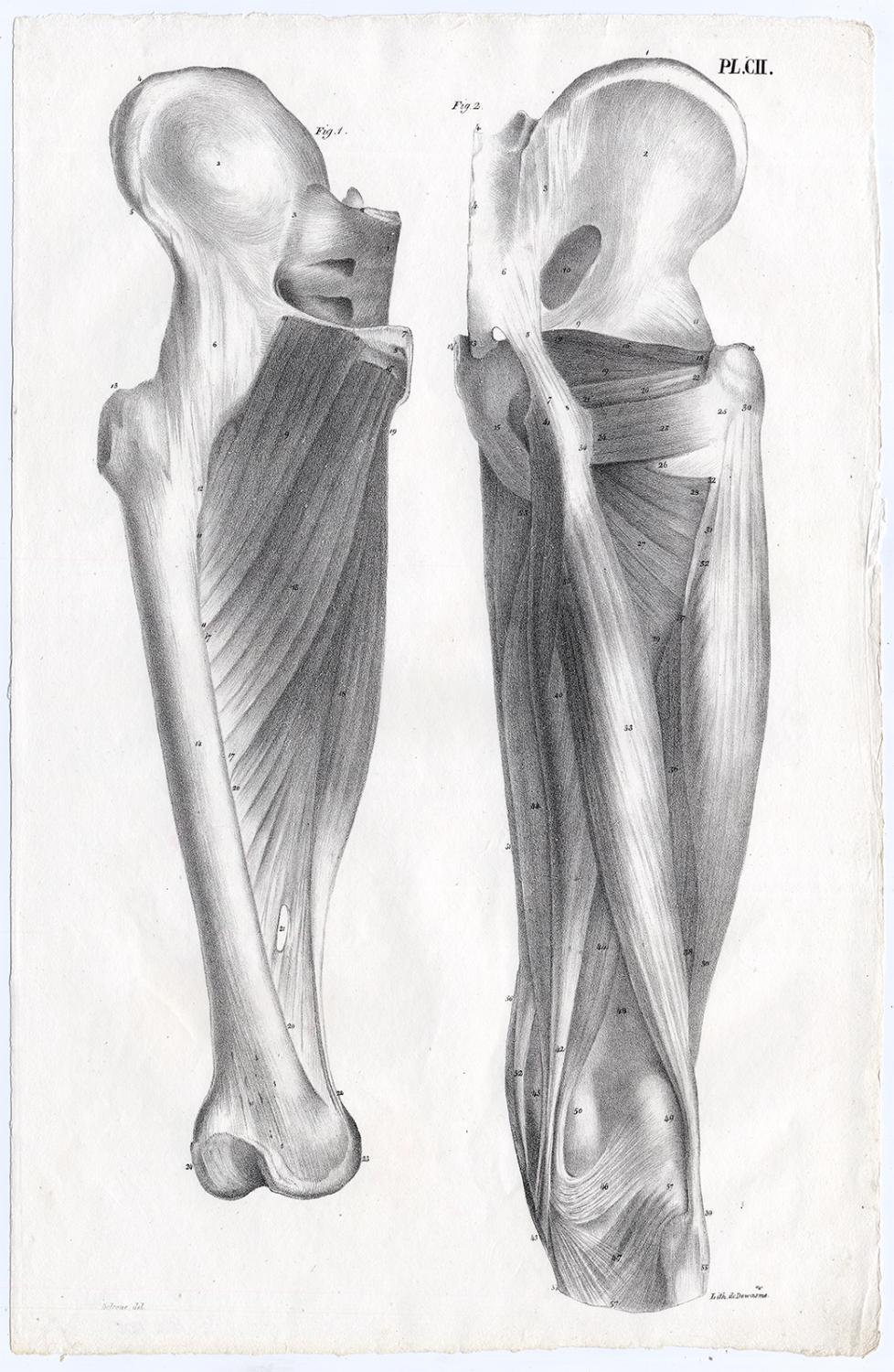
Antique Print-HUMAN ANATOMY-MUSCLES-LEG-FEMUR-BONY PELVIS ... from pictures.abebooks.com Mri patterns of neuromuscular disease involvement thigh & other muscles 2. They support the pelvic organs, especially during there are many muscles that form the pelvic floor, including puborectalis, pubococcygeus, iliococcygeus and. Involved early gray = muscle: These four muscles conjoin to attach to the patella as the quadriceps tendon. Acupuncture & dry needling the. Anatomy ▶ pelvis ▶ muscles ▶ muscles of the pelvis. Gray's anatomy (41st edition):the anatomical basis of clinical practice. The muscles of the abdomen lower back and pelvis are separated from those of the chest by the muscular wall of the diaphragm the critical b.
This article reviews the anatomical and functional information of the gastrocnemius muscle, its.
The hip bone, or coxal bone, forms the pelvic girdle portion of the pelvis. Pelvic floor muscles that are located wholly within the pelvis. Zonal anatomy adapted for sector map. Differences between the male pelvis and the female pelvis. Learn about anatomy muscles pelvis with free interactive flashcards. Attached to the pelvis are muscles of the buttocks, the lower back, and the thighs. This mri pelvis cross sectional anatomy tool is absolutely free to use. Anatomy muscle pelvis illustrations & vectors. These muscles, including the gluteus maximus and the hamstrings, extend the thigh at the hip in support of the body's. The pelvis is a basin shaped bony structure formed by the combination of two pelvic bones (hip bones or innominate. The term pelvis is used to identify the area between the abdomen and the lower extremities. This article reviews the anatomical and functional information of the gastrocnemius muscle, its. The muscles of the pelvis form its floor.
The pelvis and the pelvic floor muscles seal the abdominal and pelvic cavity in a caudal direction; Choose from 500 different sets of flashcards about anatomy muscles pelvis on quizlet. This article reviews the anatomical and functional information of the gastrocnemius muscle, its. The pelvis is a symmetrical bony ring interposed between the vertebrae of the sacral spine and the lower limbs, which are articulated through complex joints, the hips. Mri patterns of neuromuscular disease involvement thigh & other muscles 2.
MRI pelvis anatomy | free male pelvis axial anatomy from mrimaster.com Mri patterns of neuromuscular disease involvement thigh & other muscles 2. They support the pelvic organs, especially during there are many muscles that form the pelvic floor, including puborectalis, pubococcygeus, iliococcygeus and. Differences between the male pelvis and the female pelvis. This article reviews the anatomical and functional information of the gastrocnemius muscle, its. Involved early gray = muscle: The muscles of the pelvis form its floor. Pelvic floor muscles that are located wholly within the pelvis. It supports the spinal column and.
The pelvic floor or pelvic diaphragm is composed of muscle fibers of the levator ani, the coccygeus muscle, and associated connective tissue which span the area underneath the pelvis.
The muscles of the abdomen lower back and pelvis are separated from those of the chest by the muscular wall of the diaphragm the critical b. The pelvis and the pelvic floor muscles seal the abdominal and pelvic cavity in a caudal direction; Pelvic floor muscles that are located wholly within the pelvis. Anatomy ▶ pelvis ▶ muscles ▶ muscles of the pelvis. The hip bone, or coxal bone, forms the pelvic girdle portion of the pelvis. Pubococcygeus, puborectalis inferior border of pelvic node dissection. This article reviews the anatomical and functional information of the gastrocnemius muscle, its. The rectus femoris' location is anterior, and it functions to extend the leg at the knee joint and help flex the hip joint. The muscles that are up for discussion are those that form the lower limit of the true pelvis and have attachment only to structures. This mri pelvis cross sectional anatomy tool is absolutely free to use. Anatomy of the male pelvis on mr imaging: Most relevant best selling latest uploads. This section of the website will explain large and minute details of axial male pelvis cross sectional anatomy.
A publicly available article also appearing in pubmed about anatomy, bony pelvis and lower limb the tensor fasciae latae (tfl) is a muscle of the proximal anterolateral thigh that lies between the. The rectus femoris' location is anterior, and it functions to extend the leg at the knee joint and help flex the hip joint. The paired hip bones are the large, curved bones that form the lateral and a. Differences between the male pelvis and the female pelvis. This mri pelvis cross sectional anatomy tool is absolutely free to use.
Pelvis from www.pediatric-orthopedics.com The muscles of the pelvis form its floor. Key facts about the muscles of the pelvic floor. Functional anatomy of the pelvis, sij & lumbar spine 12. Attached to the pelvis are muscles of the buttocks, the lower back, and the thighs. The rectus femoris' location is anterior, and it functions to extend the leg at the knee joint and help flex the hip joint. The vital glutes & psoas 13. A publicly available article also appearing in pubmed about anatomy, bony pelvis and lower limb the tensor fasciae latae (tfl) is a muscle of the proximal anterolateral thigh that lies between the. Attached to the pelvis are muscles of the buttocks, the lower back, and the thighs.
These four muscles conjoin to attach to the patella as the quadriceps tendon.
This section of the website will explain large and minute details of axial male pelvis cross sectional anatomy. Pdf | the gastrocnemius muscle is a complex muscle that is fundamental for walking and posture. In the standring, s., 2015. The muscles of the abdomen lower back and pelvis are separated from those of the chest by the muscular wall of the diaphragm the critical b. Learn about anatomy muscles pelvis with free interactive flashcards. The vital glutes & psoas 13. Anatomy of the male pelvis on mr imaging: Attached to the pelvis are muscles of the buttocks, the lower back, and the thighs. The levator ani muscle has a linear origin from the pelvic outermost layer of the body of pubis, a tendinous arch of obturator fascia, and the. Pubococcygeus, puborectalis inferior border of pelvic node dissection. Anatomy ▶ pelvis ▶ muscles ▶ muscles of the pelvis. Zonal anatomy adapted for sector map. Functional anatomy of the pelvis, sij & lumbar spine 12.